X-rays are a form of high-energy electromagnetic radiation with the ability to penetrate most materials, making them indispensable in medical diagnostics, industrial testing, scientific research, and security screening. Their discovery in the late 19th century revolutionized multiple fields, and continuous advancements have further improved their applications. This document provides an in-depth study of X-rays, covering their discovery, generation, specificity, types, development, qualities, and uses, with detailed explanations and examples.
Discovery of X-Rays
Wilhelm Röntgen’s Experiment (1895)
- X-rays were discovered on November 8, 1895, by Wilhelm Conrad Röntgen, a German physicist, during an experiment with cathode ray tubes (early vacuum tubes used to study electron flow). He noticed that:
- A fluorescent screen in his lab started glowing, even though it was not in direct contact with the cathode rays.
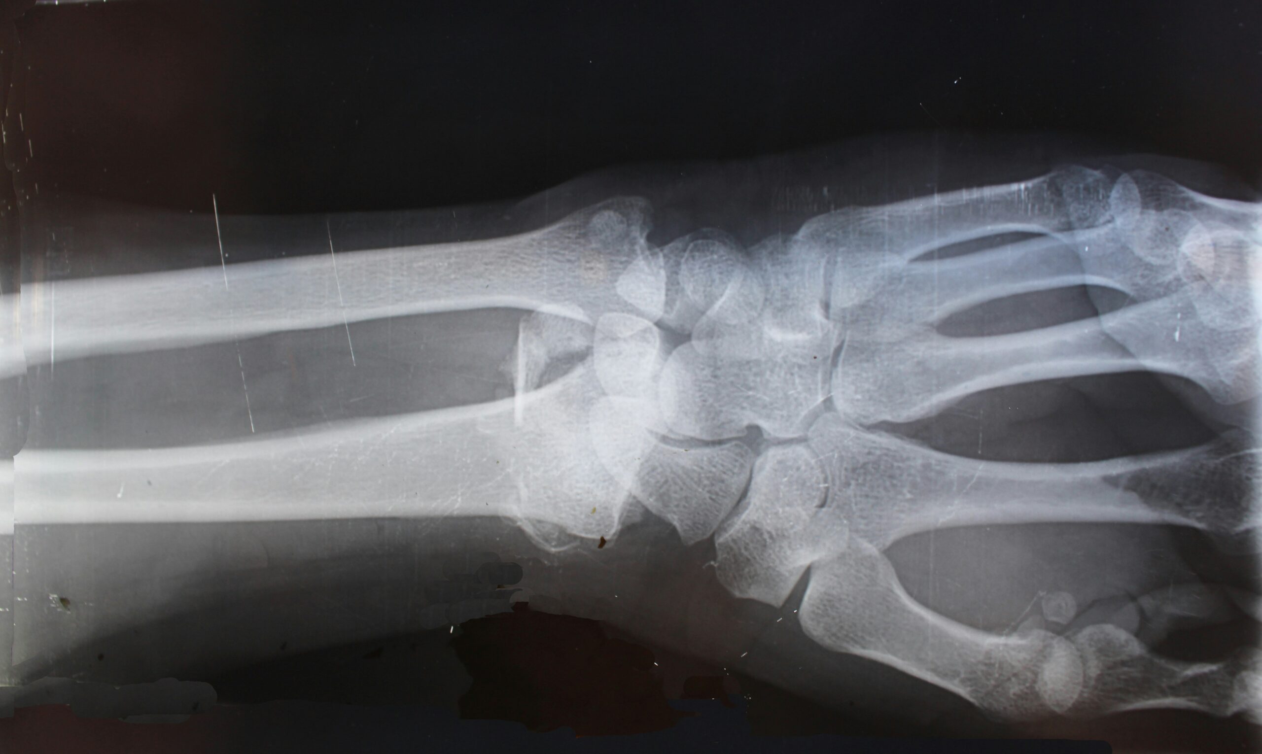
- The radiation could pass through paper, wood, and human tissues, but was blocked by denser materials like bones and metal objects.
- He captured the first X-ray image, showing the bones of his wife Anna Bertha Ludwig’s hand, along with her wedding ring.
Early Applications (1895–1900s)
- 1896: Thomas Edison and Clarence Dally experimented with X-rays for medical imaging but also discovered their harmful effects when Dally developed severe radiation burns.
- 1897: X-rays were used in the Balkan Wars to locate bullets and shrapnel inside wounded soldiers.
- 1901: Wilhelm Röntgen was awarded the first Nobel Prize in Physics for his discovery.
Generation of X-Rays
X-rays are produced when high-speed electrons strike a metal target, leading to the emission of electromagnetic radiation. This occurs inside an X-ray tube, the main component of X-ray machines.
A. Structure of an X-Ray Tube
- Cathode (- Terminal): A heated filament that emits electrons via thermionic emission.
- Anode (+ Terminal): Made of tungsten (W), molybdenum (Mo), or rhodium (Rh), which absorbs electrons and produces X-rays.
- Vacuum Chamber: Prevents interference from air molecules.
- High-Voltage Supply: Accelerates electrons toward the anode, generating X-rays upon impact.
B. Types of X-Ray Production
- Bremsstrahlung (Braking Radiation):
- Occurs when high-speed electrons slow down upon colliding with the nucleus of target atoms.
- Produces a continuous spectrum of X-ray wavelengths.
- Example: Used in general radiography (chest X-rays, bone imaging).
- Characteristic X-Radiation:
- Occurs when an inner-shell electron is ejected, and an electron from a higher shell falls into its place, emitting X-rays with specific wavelengths.
- Example: Used in X-ray fluorescence (XRF) spectroscopy for material analysis.
Specificity of X-Rays
A. Unique Properties of X-Rays
- Penetration Ability: X-rays pass through soft tissues but are absorbed by denser materials like bones and metals.
- Ionizing Power: X-rays can remove electrons from atoms, leading to chemical and biological changes.
- No Reflection or Refraction: Unlike visible light, X-rays do not reflect or refract easily.
- Fluorescence Induction: Certain materials emit visible light when exposed to X-rays, used in X-ray screens and security scanners.
B. Wavelength and Energy Range
- Wavelength: 0.01 nm – 10 nm (shorter than visible light).
- Energy: 100 eV – 100 keV (higher energy than ultraviolet radiation).
Types of X-Rays
- Soft X-Rays (0.1–10 keV):
- Lower penetration power, easily absorbed by air.
- Used in biological imaging, soft tissue scans, and X-ray microscopy.
- Hard X-Rays (10–100 keV):
- Can penetrate bones, metals, and dense structures.
- Used in medical diagnostics, industrial testing, and security screening.
- Diagnostic X-Rays:
- Low-dose radiation used in medical imaging (X-ray radiographs, CT scans).
- Example: Chest X-rays for pneumonia detection.
- Therapeutic X-Rays (Radiation Therapy):
- High-dose targeted X-rays used to destroy cancer cells.
- Example: X-ray therapy in oncology treatments.
- Industrial X-Rays:
- Used for non-destructive testing (NDT) to inspect materials without damage.
- Example: Weld inspection in aircraft and bridge construction.
Development of X-Ray Technology
- 1913: Coolidge Tube (William Coolidge) improved efficiency and image clarity.
- 1967: Computed Tomography (CT) Scan invented by Godfrey Hounsfield, producing cross-sectional images.
- 2000s–Present: Digital X-ray (DR) replaced film-based imaging, reducing radiation exposure and improving image quality.
- Future Trends: AI-enhanced diagnostics, portable X-ray devices, and nanotechnology-based X-ray imaging.
Qualities of X-Rays
- High Penetration Power – Passes through soft tissues, absorbed by dense materials.
- Ionization Effect – Can alter cellular DNA, used in cancer treatment but also poses risks.
- Fluorescent Property – Helps in X-ray fluorescence spectroscopy.
- No Charge or Mass – Behaves like light but with much higher energy.
Uses of X-Rays
A. Medical Applications
- Fracture Detection: Bone injuries are diagnosed via X-ray radiographs.
- Lung Diseases: X-rays detect pneumonia, tuberculosis, and lung cancer.
- CT Scans: 3D imaging for internal organ examination.
- Mammography: Early detection of breast cancer.
- Dental Radiography: Diagnoses cavities, root infections, and jaw issues.
B. Industrial Applications
- Non-Destructive Testing (NDT): Checks for weld defects, corrosion, and structural integrity.
- Security Scanning: Used at airports to detect contraband and explosives.
C. Scientific Research
- X-Ray Crystallography: Determines the structure of molecules, e.g., DNA (Rosalind Franklin’s discovery).
- Synchrotron Radiation: Produces high-energy X-rays for advanced physics research.
D. Environmental & Forensic Uses
- Archaeology: Analyzes ancient fossils, mummies, and paintings.
- Forensic Science: Examines bullet wounds, fake documents, and crime scene evidence.
Important Facts About X-Rays
- Wilhelm Röntgen refused to patent X-rays, ensuring free access for medical use.
- Roentgenium (Rg), element 111, was named in Röntgen’s honor.
- NASA uses X-ray telescopes to study black holes and neutron stars.
- Excess X-ray exposure can cause radiation sickness, cancer, and genetic mutations.
Conclusion
X-rays have transformed medicine, industry, research, and security with their ability to penetrate materials and reveal internal structures. Advancements in digital imaging, AI diagnostics, and nanotechnology continue to expand their applications, making them an irreplaceable tool in modern science and technology.